
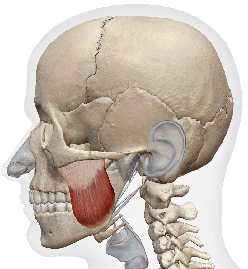
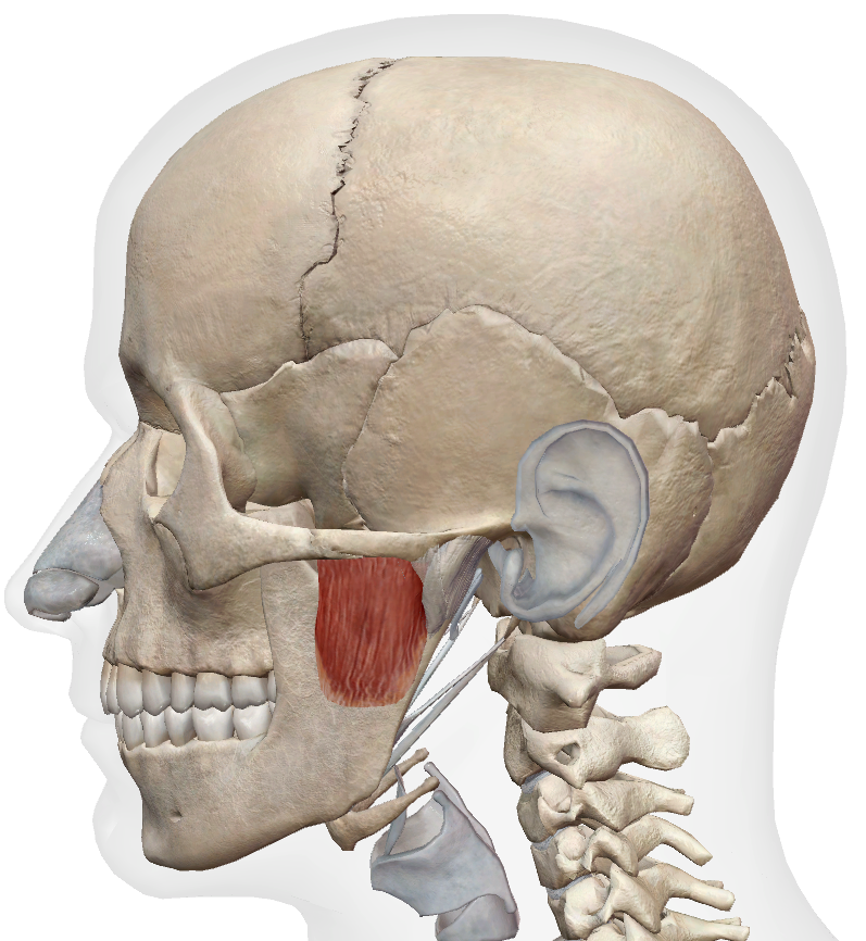
Masseters
Superficial & Deep heads · See Scenario 1: TMD

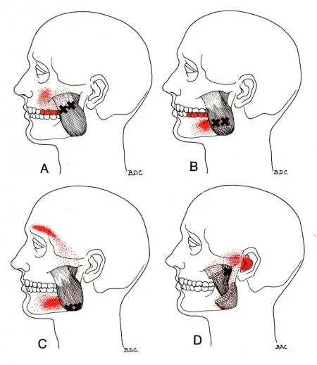
| Superficial head | Lower molars, cheek, lower jaw — often mistaken for dental pain |
| Deep head | Ear, TMJ region, zygoma — can mimic ear pain or joint dysfunction |
Client position: Supine (preferred) or seated. Jaw slightly open and fully relaxed — teeth apart, lips lightly touching.
Client communication: “I’m going to work along the side of your jaw. The area can be quite tender, especially if you clench. Breathe normally. Let me know if the pressure is too strong and I’ll ease off.”
Locate & confirm: Ask the client to clench gently — the superficial belly hardens and bulges prominently over the ramus and angle of the jaw. Ask them to relax and confirm softening. The transition between contracted and relaxed helps you map the muscle boundary.
Superficial head
Palpate: Broad belly over the lateral mandibular ramus, from the angle of the jaw upward toward the zygomatic arch. Work in sections — inferior (near the mandibular angle), mid-belly, and superior (below the zygomatic arch). Compare sides.
What to feel for: Diffuse hypertonicity across the belly (very common in bruxers), taut bands particularly in the inferior and mid sections, tenderness that reproduces jaw ache or molar pain, asymmetry between sides.
Deep head
Palpate: Fingertip approach just anterior to the tragus of the ear, above the zygomatic arch. Requires firmer, more directed pressure in a slightly posterior direction. Ask client to open and close very slightly — the deep head shifts under the finger on movement.
What to feel for: Deep, localised tenderness reproducing ear pain or TMJ region pain — confirm verbally. Less diffuse than the superficial head; a focused nodule is more typical.
Superficial head: Pin the belly with thumb or flat fingertips across the fibre direction. Work from the mandibular angle upward in 3–4 sections. At each pin, ask the client to open the jaw slowly and fully, then return to neutral. Maintain pin pressure throughout. Repeat 4–6 cycles per section. Adjust pin location between passes to cover the full belly.
Deep head: Fingertip pin just anterior to the tragus, directed slightly posteriorly. Ask the client to open the jaw slowly, then return to neutral. 4–6 cycles. The range of movement available here is smaller — controlled, partial opening is sufficient.
Superficial head: Work systematically across the belly in sections. Sustained pressure on identified taut bands or nodules — firm but within tolerable range. Hold 60–90 seconds or until softening and referred pain reduction. Referral to the lower molars during treatment is common and diagnostically useful — confirm it reproduces familiar symptoms. Maximum 2–3 trigger points per session.
Deep head: Sustained fingertip pressure anterior to the tragus. Hold 60–90 seconds. Do not increase pressure if strong ear or joint-adjacent pain is present — maintain and wait for release.
The masseter is a jaw closer. PIR is the appropriate approach: resisted jaw closing at end of opening range produces post-isometric relaxation and passive gain in opening.
Client supine. Bring the jaw to end of comfortable opening range. Place a cupped hand under the chin. Ask the client to close the jaw gently against your upward resistance (10–20% effort). Hold 5–7 seconds. Relax — jaw drops open passively, do not force it. Repeat 3–5 times.
Cross-fibre strokes at the inferior attachment near the angle of the mandible where fascial adhesions or thickening are palpable. Firm and precise — 30–60 seconds. Not a primary technique; use when the attachment is specifically reactive and superficial belly work alone is insufficient.
Parotid gland: The parotid gland sits directly posterior to the masseter. Deep pressure behind the posterior border of the muscle risks compressing the gland. Stay anterior to the posterior ramus border when working the deep head. If the client reports a sudden increase in jaw or throat discomfort, reposition anteriorly before continuing.
Facial nerve: The facial nerve (CN VII) exits the parotid gland and branches across the face in the posterior masseter region. Avoid sustained deep pressure behind the posterior ramus border. Work the deep head from an anterior approach only.
Pressure threshold: The masseter can be extremely tender in chronic clenchers. Start light and build gradually — confirm tolerance before increasing depth. If a headache starts or worsens during treatment, reduce pressure and reassess before continuing.
Technique limit: If jaw symptoms worsen, TMJ clicking increases, or the client reports sharp or catching pain during treatment — stop and reassess. Do not push through sharp joint-adjacent pain.